Written by Amanda Callaghan, Curator/Director of the Cole Museum of Zoology at the University of Reading.
The narwhal Monodon monoceros is one of the rarest and oddest-looking whales. Cousin to the white Beluga whale, narwhal means “corpse whale” in Icelandic, (nar corpse and hvalr whale) a reference to its blotchy grey pallor1. Male narwhals (and a small number of females) have a canine tooth on the left-hand side of the upper jaw that grows through the upper lip into a long spiral “tusk.” The function of this strange tooth is unclear, but the horns are used for duelling or ‘tusking’ when the males compete for females.
From medieval times through to the 17th century, the legend of the mythical horned horse, or unicorn, was reinforced by the spectacular spiralling horns brought to Europe. Those observed live in the seas around Greenland and Iceland were known as “sea unicorns”2. Today, although we know they are not unicorn horns, there remains a fascination and high price for these rare and beautiful teeth.
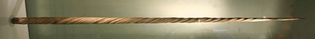
We are lucky to have five accessioned narwhal specimens in the Cole Museum. The first is a 2m long narwhal tusk of unknown provenance, donated to the Cole Museum in 1943 by William A. Smallcombe who was the curator of Reading Museum between 1926 and 19573 (Fig 1).
The remainder are thanks to the research of former curator Dr Nellie B. Eales who wrote two papers on foetal narwhals, one on the skull and the other on the manus 4,5. The skull work was illustrated with her line drawings and several 3D models made using the wax plate method6.
Eales obtained the foetuses from geologist Professor L. R. Wager, a one-time Lecturer in Petrology at the University of Reading. He left his Reading post after 6 months to join an expedition to Greenland, and went on to lead the Courtauld Expedition to Kangerlussuaq Fjord7. Following a request from the formidable Dr Eales, he returned with three narwhal foetuses for her to study. These would have been obtained by the local Inuit people and Eales noted in her paper that they gifted narwhal foetuses to their children as baby dolls, swaddled in material4.
What remains of the narwhal foetuses?
On their arrival to England the narwhal foetuses, swaddled like babies, were designated A, B and C. Foetus A, female, (137 mm long, 30 mm broad, 32 mm high) was sectioned and used for the skull model (Fig. 4).
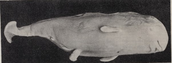
Foetus B, female, (150 mm long, 24 mm broad and 40 mm high), distorted by poor packaging, was used to study the head but no model seems to have been made. Specimens A and B were destroyed during the research procedure. Foetus C, (280 mm long, 60 mm broad, and 70 mm high) was not used as it was “too developed.” The specimen in figure 3, which matches this description and resembles foetus A, is probably foetus C, but it is odd that it was not accessioned into the museum.
The narwhal models.
Three narwhal models were made with the state-of-the-art wax plate method. The head of foetus A was cut off and embedded in celloidin for sectioning by Dr Keith Richardson of University College, London. The skull sections were reproduced using an Edinger projector and a Zeiss planar lens to magnify the image4. These were transferred to 1mm beeswax plates and painted (example in fig. 4).
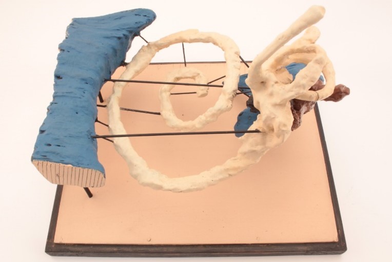
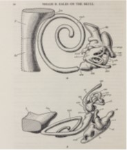
Figure 4. Narwhal skull model COLE3061. Left: Reconstructed wax plate model of inner ear 25x. Right: illustration of the model from Eales4.
Nellie’s paper was groundbreaking when published and her work is still relevant, cited in studies of cetacean foetal skull morphology8. Thankfully, although they are frail, the models have survived the past 75 years despite being housed for many years in a dusty and overheated teaching laboratory. They will be moved to a new storage facility in 2024 on shelving that allows for easy viewing of these historic models.
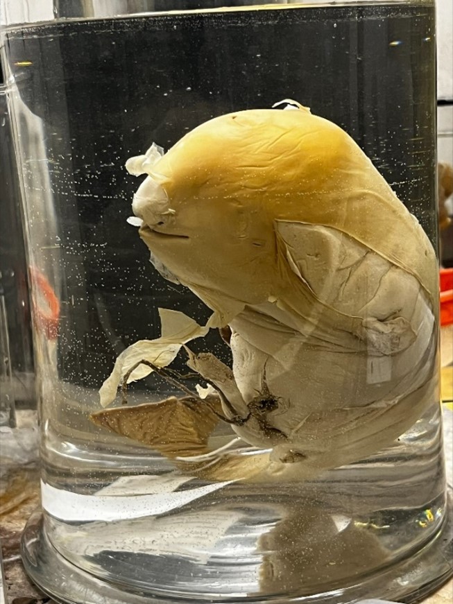
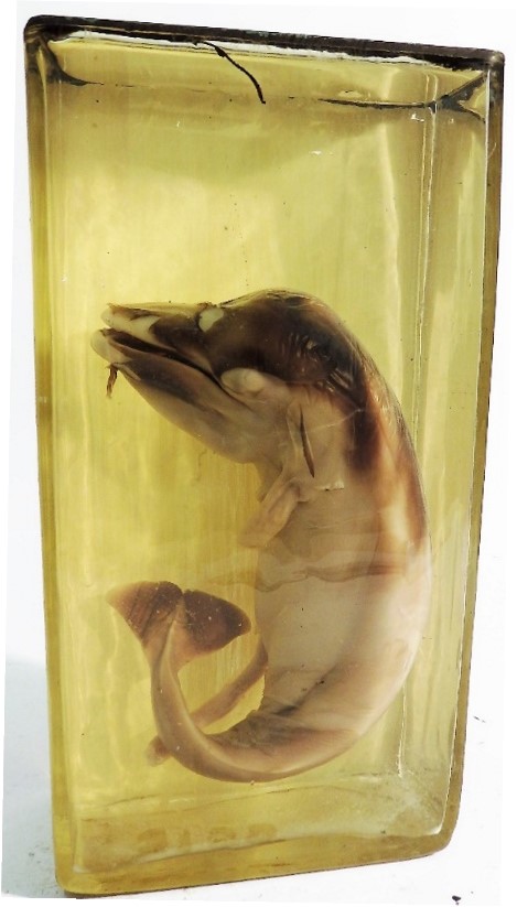
The fifth and final narwhal specimen is an odd one. We have a “narwhal foetus” (COLE3156) accessioned in 1953, not long after the narwhal research, by new curator Bill Stoneman (Fig. 5). A consultation with Dr Agneze Lanzetti from the Natural History Museum in London has led us to believe that COLE3156 is a baleen whale of the genus Balaenoptera.
Scientists continue to publish descriptive morphological studies of foetal cetaceans, albeit with more sophisticated visualisation, including CT scans and 3D computer modelling9. We are hoping that we will soon be able to CT scan both our cetacean foetuses to confirm their identification, correct the catalogue, continue Dr Eales research, and add to the collection with some modern 3D models.
References
- Britannica, The Editors of Encyclopaedia. “narwhal”. Encyclopedia Britannica, 25 Feb. 2024, https://www.britannica.com/animal/narwhal. Accessed 25 March 2024.
- Dectot, X (2018). When ivory came from the seas. On some traits of the trade of raw and carved sea-mammal ivories in the Middle Ages, in Jacquemard C., Gauvin B., Lucas-Avenel M.-A., Clavel B. & Buquet T. (éds), Animaux aquatiques et monstres des mers septentrionales (imaginer, connaître, exploiter, de l’Antiquité à 1600). Anthropozoologica 53 (14): 159-174.
- Reading Museum 2024. William Smallcombe, curator at Reading Museum. http://collections.readingmuseum.org.uk Accessed 13/03/2024.
- Eales, N.B. (1950). The skull of the foetal narwhal, Monodon monoceros L. Philosophical Transactions of the Royal Society of London, Series B. Biological Sciences, 612(235), pp 1-33.
- Eales, N.B. (1954). The manus of the narwhal Monodon monoceros. L. Proceedings of the Zoological Society of London, vol. 124: 201-211.
- Hopwood, N. (1999). ” Giving Body” to Embryos: Modelling, Mechanism, and the Microtome in Late Nineteenth-Century Anatomy. Isis, 90(3), 462-496.
- Wager, L.R., Deer, W.A., Wager, H.G. and Manley, G., 1937. The Kangerlussuaq Region of East Greenland. The Geographical Journal, 90(5), pp.393-421.
- Nweeia, M.T., Eichmiller, F.C., Hauschka, P.V., Tyler, E., Mead, J.G., Potter, C.W., Angnatsiak, D.P., Richard, P.R., Orr, J.R. and Black, S.R., 2012. Vestigial tooth anatomy and tusk nomenclature for Monodon monoceros. The Anatomical Record: Advances in Integrative Anatomy and Evolutionary Biology, 295(6), pp.1006-1016.
- Rauschmann, M.A., Huggenberger, S., Kossatz, L.S. and Oelschlaeger, H.H., 2006. Head morphology in perinatal dolphins: a window into phylogeny and ontogeny. Journal of Morphology, 267(11), pp.1295-1315.


Pingback: NatSCA Digital Digest – June 2024 | NatSCA