Written by Claire Kelly, Conservator at Natural History Museum, London.
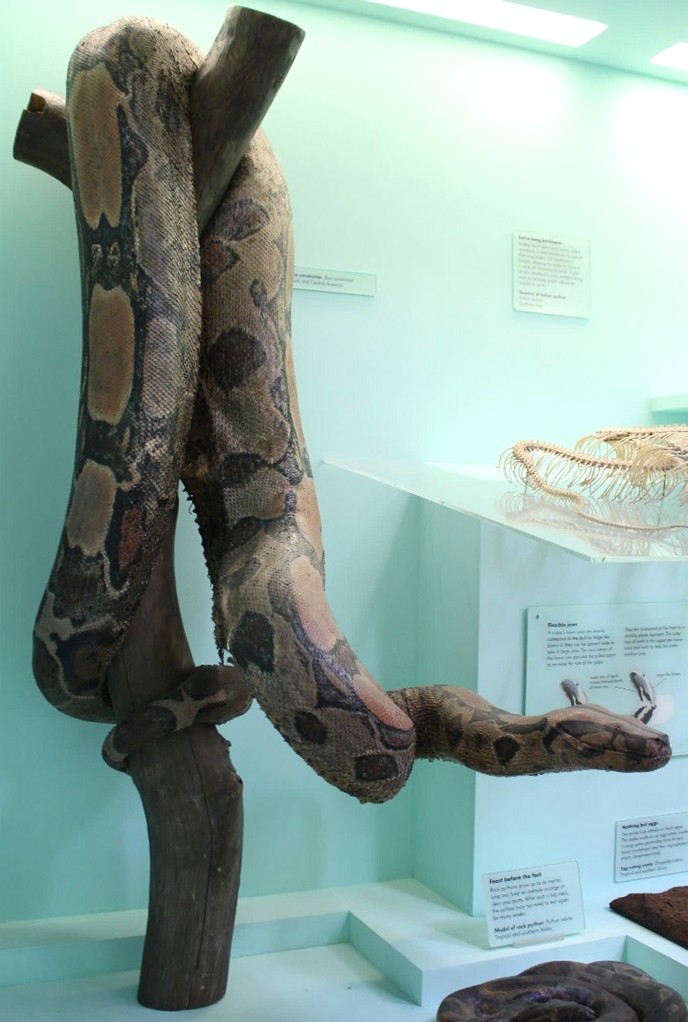
A Boa Constrictor on display in the Fishes, Amphibians and Reptiles Gallery at the Natural History Museum in London, UK was removed to undergo a considerable amount of remedial conservation treatment.
The taxidermy skin, dating from around the late 19th century, is mounted over a plaster form that was placed onto a wooden trunk. The skin exhibited severe deterioration with multiple splits in various areas located throughout the length of the specimen. The entire ventral seam had opened along with skin distortion and lifting around the splits without any stitched seam to hold it in situ. Most of the damage was at the ventral area of the specimen but some splits and distortion were visible whilst on display, along with material shed on to the case base.
The current display case was created around 1994; it has poor environmental conditions that can vary greatly in terms of humidity and temperature, this is not to the standard of display cases that the museum would install today. The case is housed in a closed ended corridor. It is part of the gallery corridor internal walls with apertures cut into the wall, forming part of a display of 12 similar structures. They all consist of a wooden box structure constructed within the walls, with a large glass frontage set in a wooden frame. This glass frontage must be removed to access the case and the specimens.
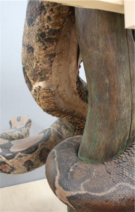
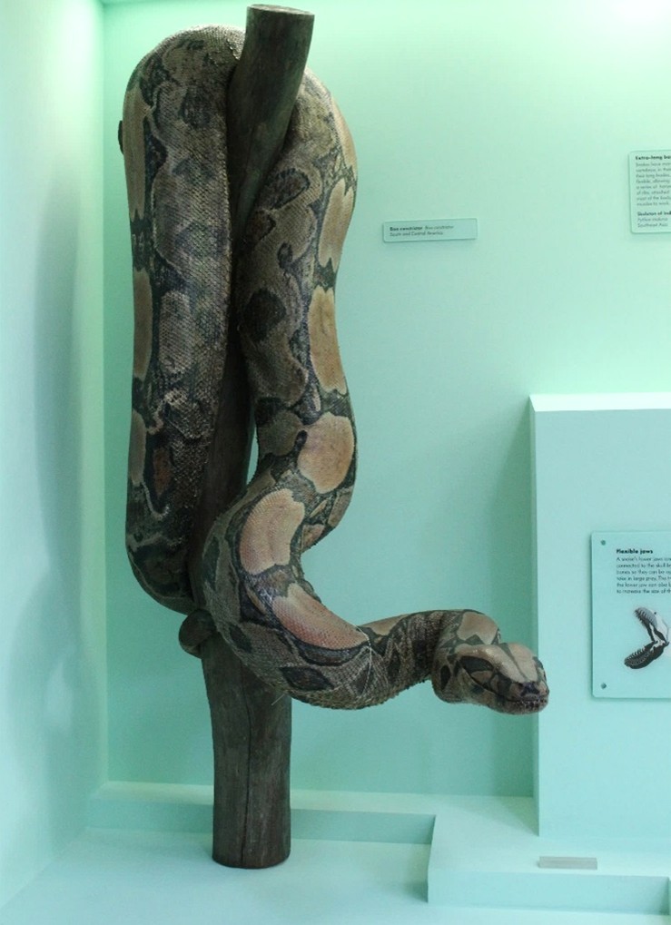
The awkward shape of the specimen with its protruding head and 1.5m height made access for treatment quite difficult. A wooden skate with a minimal wood frame was fabricated to allow safe and easy movement whilst maximising access for treatment and maintaining the vertical position.
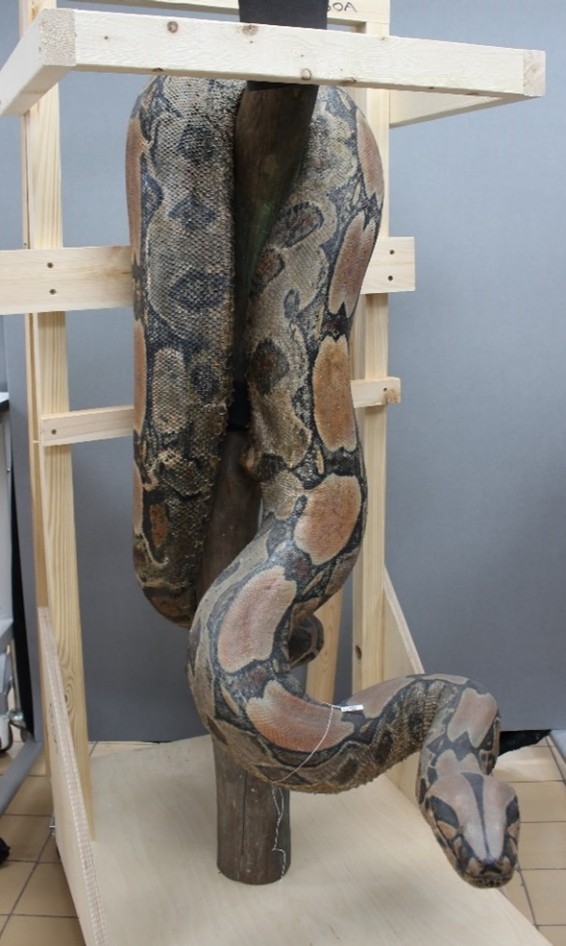
Boa Constrictor on a bespoke minimal wheeled frame prior to remedial treatment ©The Trustees of the Natural History Museum
An initial assessment revealed that the skin may have receded at some point as the seams appear to have been severed. It is possible that when it was tanned and placed on the form it started to shrink and recede causing the preparator to cut the seams to prevent the skin from splitting, this may be the cause of the initial splits. The inside of the skin also displays plaster residue from when it must have originally been positioned on to a soft plaster surface.
The skin is heavily overpainted, presumably a historic decision for aesthetic purposes to enhance the colours of the skin, as is seen in other display specimens that demonstrate loss of depth of colour after death.
The skin was tested using a portable XRF for the presence of any contaminants and showed a high concentration of lead. This may be from the paint used to enhance the aesthetic appearance.
The whole specimen was carefully dry cleaned with a soft brush and vacuum, cosmetic sponge, washed prior to use, cut into fronds to prevent damage to scale. Smoke sponge was used on more robust areas which were then vacuumed again to remove any residue.
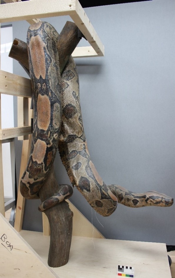
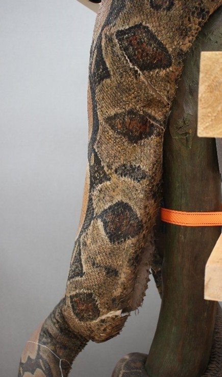
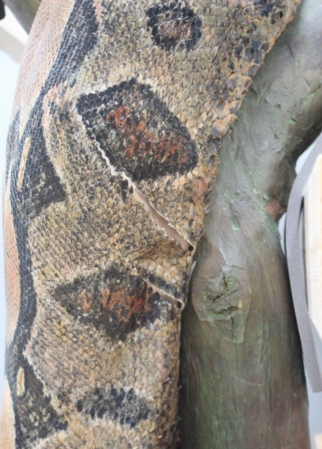
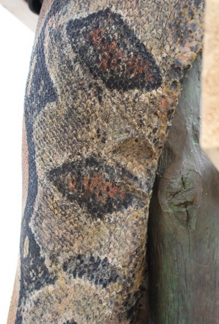
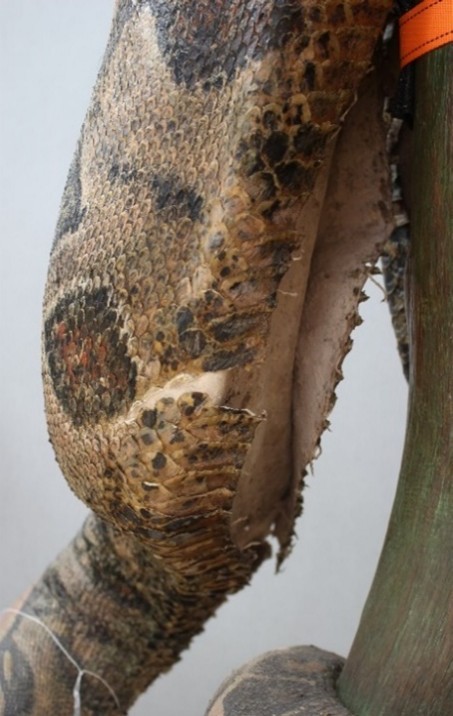
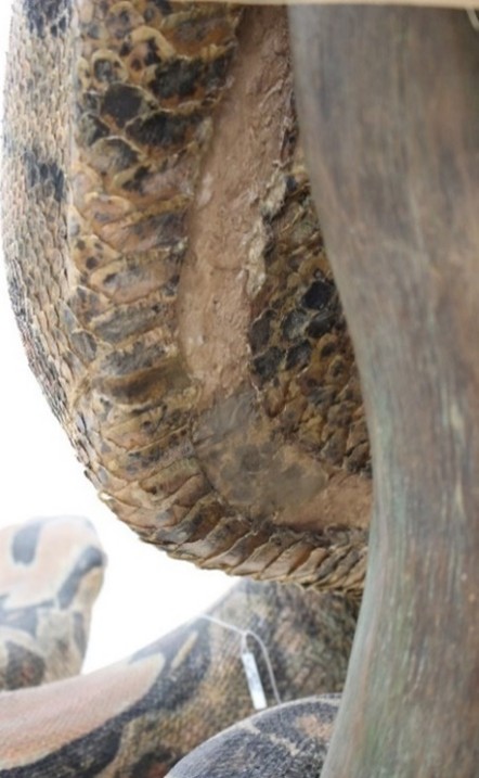
Most of the damaged areas were splits and distortion originating from the seam. These were mainly located in areas that had very limited access due to the proximity between the ventral surface of the seams and the wooden branch support.
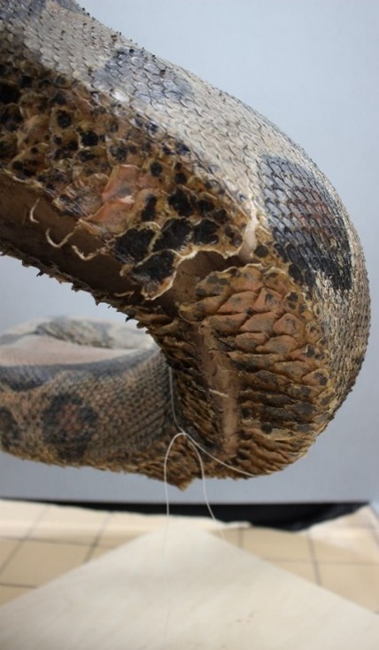
Examples of distortion and splits in the skin ©The Trustees of the Natural History Museum
The distortion of the skin was rectified using localised humidification. After initial testing with a humidifier, it soon became clear this was not a suitable methodology due to the thickness of the skin and scales. Their thickness required a very long dwell time and also there was a lack of control of the mist onto a plaster surface. Further tests were carried out using a method based on one for label removal. A blotter sandwich – small sections of acid free blotting paper were wetted using deionised water, then placed together and positioned over Bondina™ (non-woven polyester) larger than the size of the blotter, this sandwich of materials was then required to be held in-situ against the vertical surface of the distorted skin.
Due to the awkward positioning of the damaged and distorted areas, it required a little creative thinking to 1. Ensure the humidification sandwich was held in contact with the skin on a vertical surface or underside surface and 2. to maintain a reasonable amount of pressure to realign.
A novel approach was devised using clingfilm, cotton tape and pads of Plastazote™ (closed-cell, crosslinked polyethylene (XLPE) foam). The Plastazote™ pad was held in place with a slot cut into the foam structure and cotton tape was fed into the slot with enough tape to wrap around the snake. Clingfilm was wrapped over the humidifying sandwich to contain the moisture and wrapped onto itself around the circumference of the snake. The Plastazote™ pad was then positioned over the sandwich and the tape wrapped around the circumference of the snake, over the clingfilm and could then be tied securely into position. Additional pressure/stabilisation was added where necessary by packing out with extra pieces of Plastazote™ gently pressed into the void between the snakeskin and the tree trunk.
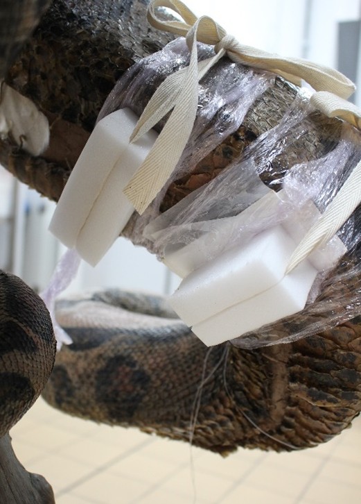
Plastazote™ pads holding the humidification sandwich in situ and tied around specimen ©The Trustees of the Natural History Museum
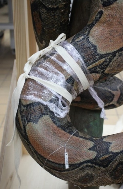
Depending on the amount of distortion and size of area affected, the rehydration was carried out in stages to initially soften and then to temporarily increase the pressure to realign, or to work gradually along a larger area.
The wetted blotter was left in place for 48 hours to allow the area to soften and realign. The packing was also left in situ to allow the area to dry to a pliable stage whilst still maintaining the pressure to keep the skin aligned. It was then possible to secure the flattened pliable surface by backing and packing the surfaces with Lascaux™ 498HV adhesive and Japanese tissue. These materials were chosen due to their sympathetic qualities for skin repairs, providing strength and some flexibility. Where splits had occurred, these areas were also backed with a stronger and heavier Japanese tissue adhered with Lascaux™ 498hv adhesive. Some areas were also initially heat set due to limited accessibility with precoated Lascaux™ 498HV Japanese tissue being inserted into the limited opening and a small, heated spatula applied onto a dry blotter pad placed over the skin to allow the heat to activate the adhesive without direct contact with the scales.
The snakeskin edges were also reinforced with the thicker Japanese tissue to help minimise any future movement and some areas were bridged with the Japanese tissue. Splits were gap filled with a layer of Lascaux™ 498HV and paper pulp mixed with acrylic paint to form a base. Pieces of Japanese tissue were then colour matched using acrylic paint and cut to form separate scales. These replicated scales were then adhered to the surface of the fills using Lascaux™ 498HV adhesive and then carefully colour matched tonally again to the surrounding area, allowing splits to be blended into the snakeskin and to disguise any visible areas of damage.
Before and After comparison images ©The Trustees of the Natural History Museum
The specimen is now stabilised and is to remain on permanent display in the Fishes, Amphibians and Reptiles gallery with regular visual inspections.


Pingback: NatSCA Digital Digest – November 2025 | NatSCA