How many natural history collections contain drawers and drawers of unloved microscope slides? With a few notable exceptions, such as the Grant Museum Micrarium, museums often find slides difficult to display and use.
The Royal College of Surgeons (RCS) has a particularly large collection of 50,000 slides, making up more than half of all the objects in the collections here. Hardly any are on display in the Hunterian Museum. A closer look however, reveals that the RCS microscope slide collections are really something special. From William Osman Hill’s Yeti slides to William Hewson’s 240 year old microscope vials, the slide collection here is every bit as exciting and important as the other objects in the museums.
As Collections Assistant for the microscope slide collection most of my work over the last six months has been on the John Thomas Quekett collection. His name is not well known, but if you have heard of it that is probably because you have come across the society of microscopists named in his honour. The Quekett Microscopical Club (QMC) has generously funded a project to care for Quekett’s original slides.
John Thomas Quekett (1815 -1861) was a leading histologist and microscopist who was Richard Owen’s deputy at the Royal College of Surgeons. Quekett took over as conservator of the Hunterian Museum in 1856 when Owen left for the British Museum to become the superintendent of the natural history department and oversee the building of what would become the Natural History Museum, London. Quekett was at the cutting edge of a revival of the popularity of the microscope in the Victorian period. He wrote A Practical Treatise on the Use of the Microscope which became a classic text for microscopists, and is known to have instructed Prince Albert in the use of his silver microscope. He was a fellow of the Linnaean Society and the Royal Society, and worked with famous scientists such as geologist Charles Lyell, palaeontologist Gideon Mantell, explorer David Livingstone, botanist Joseph Hooker and Charles Darwin himself.

John Thomas Quekett (1815-1816). (Image Royal College of Surgeons)
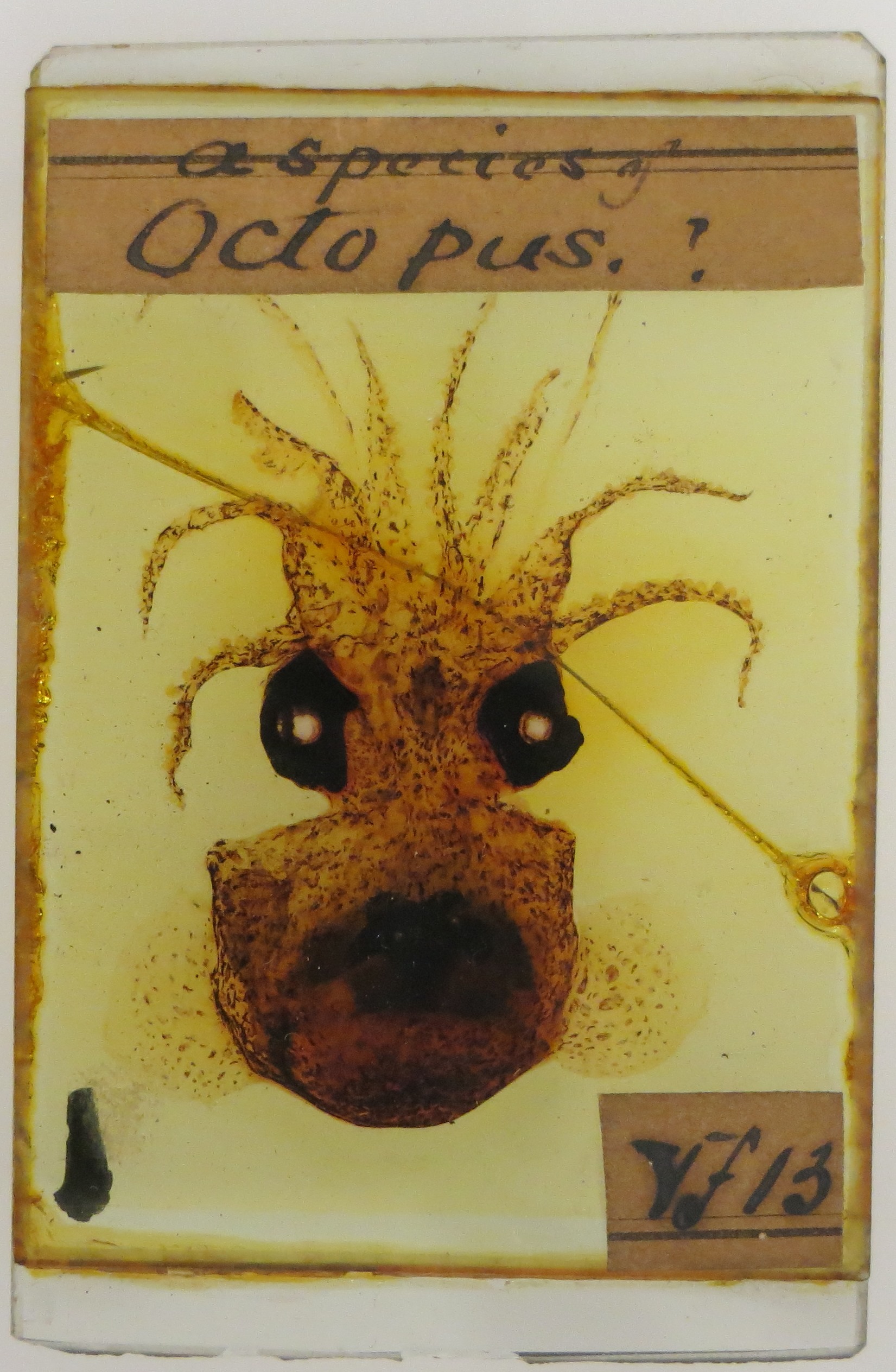
An octopus from the Quekett Microscope slide collection. (Image Royal College of Surgeons)
Quekett was a pioneer in histology and microscopy, designing his own microscope and producing stunning histological preparations, especially injected specimens. Furthermore he put every natural object he could get his hands on onto a microscope slide. Animal, vegetable, mineral, everything. Tissue samples of every organ in the human body, a whole octopus, tiny flakes of silver, the exquisitely prepared respiratory system of a caterpillar. He prepared diatoms, ferns and coal, delicate sections of pterosaur bone, thylacine teeth and oak wood. There are even hair samples disturbingly labelled ‘vampyre’, although this probably refers to the bats rather than the undead.
He carefully labelled and catalogued his slides to produce a comprehensive natural history collection on a microscopic scale, and 12,000 of these slides remain today. It is not surprising that a recent review of the RCS collections concluded that the Quekett material is “one of the strongest representative collections of Victorian microscopy and scientific practice in general in the UK and possibly the world” (RCS Significance Review June 2015).
Given their age the slides are in relatively good condition, but there are some issues to contend with such as cracked glass, missing labels and leaking fluid. Since the 1880s microscope slides have been a standard size – 2.5cm x 7.5cm, but the Quekett collection predates this. His slides range in size from 1.8cm x 4.8cm up to a whopping 8.5cm x 20cm. Some of the slides are also very thick and all this makes storage difficult and any type of automated scanning nigh on impossible.

Slices through the teeth of thylacines. (Image Royal College of Surgeons)
The collection is obviously of interest to those working on the history of science and microscopy, but impressively the slides are still being used for scientific research, 170 years after Quekett made them. Preparations of harder materials such as fossils, bones and teeth have survived in excellent condition, enabling modern researchers to gather data from the collection. The image below was taken recently using reflected light fluorescence microscopy by a PhD student studying bone remodelling in mammal species.

Transverse section of the humerus of a mountain hare. Prepared by J T Quekett, photographed by Alessandro Felder, Royal Veterinary College. Image taken at 4x magnification using reflected light fluorescence microscopy. (Image Royal College of Surgeons)
With the upcoming RCS decant and new Hunterian Museum planned for 2020 there is an opportunity to bring this collection to prominence again. We plan to include Quekett’s story in the new exhibits and make his collection better known, better protected and more easily accessible online. At first glance an old microscope slide collection might not look like much, but if you investigate further you never know what you might find.
Written by Hannah Cornish
(Collections Assistant, Royal College of Surgeons)
For more information about J T Quekett see:
https://www.rcseng.ac.uk/library/blog/quekett-and-exploration
http://www.quekett.org/about/who/history


Pingback: NatSCA Digital Digest | NatSCA
Pingback: Top Ten Most Read Blogs of 2016 | NatSCA